 | |
|
Navigation Computerized Tomography (CT) & Magnetic Resonance Imaging (MRI) Nuclear Medicine & Positron Emission Tomography (PET)
|
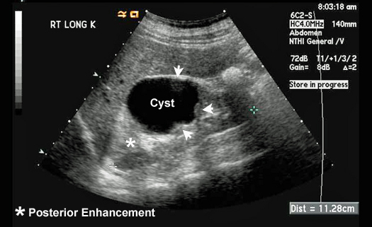
Ultrasound imaging (sonography) uses high frequency sound waves to view inside the body. Ultrasound images are captured in real-time, so they can show movement of the body's internal organs as well as blood flow through the vessels. Unlike X-ray imaging, there is no ionizing radiation exposure associated with ultrasound imaging. In an ultrasound exam, a transducer (probe) is placed directly on the skin or inside a body opening. A thin layer of gel is applied to the skin so that the ultrasound waves are transmitted from the transducer through the gel into the body. The ultrasound image is produced based on the reflection of the waves off of the body structures. The strength (amplitude) of the sound signal and the time it takes for the wave to travel through the body provide the information necessary to produce an image. Uses: abdominal ultrasound (to visualize abdominal tissues and organs), breast ultrasound (to visualize breast tissue), echocardiogram (to view the heart), fetal ultrasound (to view the fetus in pregnancy), ultrasound guided needle placement (in blood vesses or other tissues of interest). Ultrasound of a complex cyst in a kidney |
|
Jamie Mize | ENG 307T | Digital Writing | Summer 2017 | |