 | |
|
Navigation Computerized Tomography (CT) & Magnetic Resonance Imaging (MRI) Nuclear Medicine & Positron Emission Tomography (PET)
|
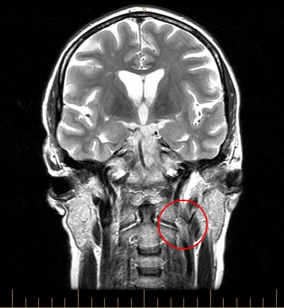
Computed tomography (CT) is a diagnostic imaging test used to create detailed images of internal organs, bones, soft tissue and blood vessels. The cross-sectional images generated during a CT scan can be reformatted in multiple planes, and can even generate three-dimensional images which can be viewed on a computer monitor, printed on film or transferred to electronic media. CT scanning is often the best method for detecting many different cancers since the images allow your doctor to confirm the presence of a tumor and determine its size and location. CT is fast, painless, noninvasive and accurate. A CT scan be used to study all parts of the body: chest, stomach, pelvis, arm, leg, liver, pancrease, kidneys, bladder, adrenal glands, lungs and heart. In some cases, a dye called contrast material will be used. This requires an IV in your arm (sometimes the contrast is given orally). This dye makes organs easier to see on the image. MRI Magnetic resonance imaging (MRI) of the body uses a powerful magnetic field, radio waves and a computer to produce detailed pictures of the inside of your body. It may be used to help diagnose or monitor treatment for a variety of conditions within the chest, abdomen and pelvis. An MRI can be done for many reasons - tumors, bleeding, injuries, blood vessel issues, or infection. an MRI can also be performed to provide additional information found on an X-ray, ultrasound or CT. Contrast material can also be used during an MRI to show abnormal tissue more clearly. An MRI scan be done on: the head, chest, blood vessels, abdomen, pelvis, bones and the spine. Head CT showing deep intracerebral hemorrhhage (also known hemorrhagic stroke) 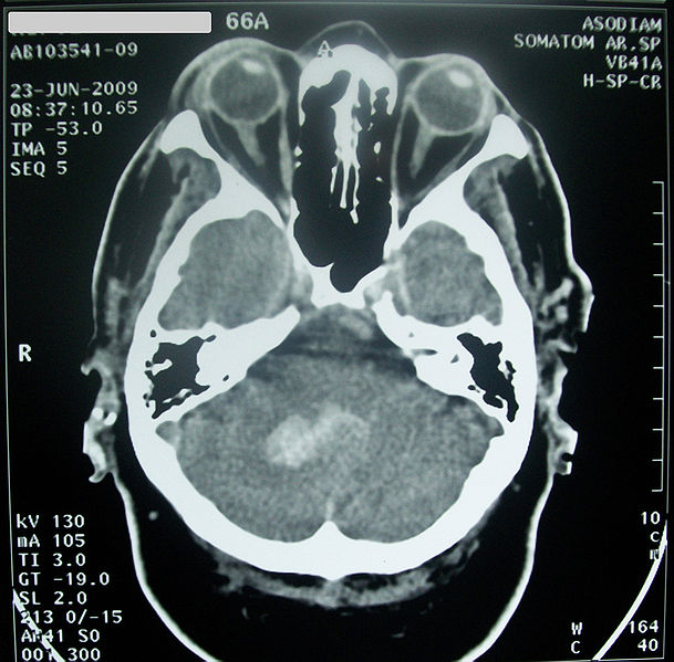 Brain/Spinal MRI |
|
Jamie Mize | ENG 307T | Digital Writing | Summer 2017 | |