 | |
|
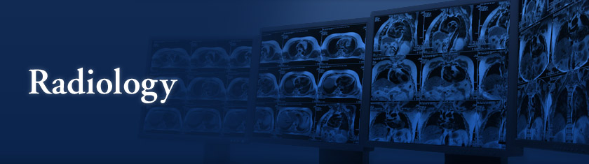
Navigation Computerized Tomography (CT) & Magnetic Resonance Imaging (MRI) Nuclear Medicine & Positron Emission Tomography (PET)
|
Nuclear medicine is a branch of medical imaging that uses small amounts of radioactive material to diagnose and determine the severity of a disease (or to treat it). This includes types of cancer, heart disease, gastrointestinal, endocrine, neurological disorders and other abnormalities that may be within the body. Nuclear medicine procedures are able to detect molecular activity, which offers the potential to identify disease in its earliest stages. Uses: to detect or visualize heart disease/issues (such as assessing heart damage after a heart attack, or detect coronary artery disease), to detect lung disease/issues (such as scaning the lungs for respiratory and blood flow problems), to evaluate and detect bone disease/issues (evaluate bones for fractures, or tumors), or to detect brain abnormalities/diseases (nuclear medicine can detect the early onset of neurological disorders such as Alzheimer's disease). Nuclear medicine imaging can also identify inflammation or abnormal gallbladder function, bleeding into the bowel, lymphedema, evaluate a fever with of unknown origin, and many, many other things.
Positron Emission Tomography (PET) is another branch of nuclear medicine imaging. It also uses small amounts of radioactive materials called radiotracers, a special camera and a computer to help evaluate your organ and tissue functions. By identifying body changes at the cellular level, PET can detect the early onset of disease before other imaging tests. PET scans, like nuclear medicine imaging, can detect and evaluate different tissue or organ diseases. It can detect heart issues, other organ issues, cancer, skin and tissue diease, etc. Unfortunately, due to the nature of this testing, there are no available images to show as an example. |
|
Jamie Mize | ENG 307T | Digital Writing | Summer 2017 | |